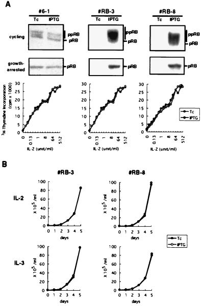
Figure 1.
Expression of pRB in cytokine-dependent hematopoietic cells. (A) Inducible expression of pRB in asynchronously growing (Top) or growth-arrested (Middle) 6–1 pro-B cells and two pRB-transfectant clones, RB-3 and RB-8, was examined by anti-pRB immunoblotting. The signals for endogenous pRB in 6–1 cells were obtained after prolonged exposure (10 times that of the other cells). Positions of the hypophosphorylated (pRB) and the hyperphosphorylated (ppRB) forms were indicated. (Bottom) The growth-arrested cells were then restimulated with various doses of IL-2 for 24 h and IL-2-dependent cell proliferation was evaluated by measuring [3H]thymidine incorporation into DNA. (B) The pRB-transfectants were cultured in medium containing IL-2 or IL-3 in the presence of Tc or IPTG, and cell numbers were counted.

