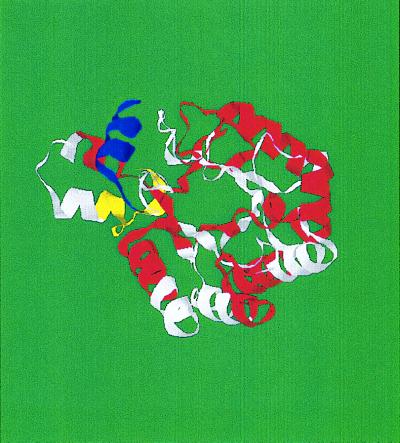
Figure 3.
Structure of the TIM-barrel protein Endo-1,4-β-d-glucanase (1TML), indicating the locations of the sequence units determined by the peak in the cosine power spectrum at k = 21. Units are alternately colored red and white, with the exception of the N-terminal unit, which is colored yellow, and the C-terminal unit, which is colored blue.

