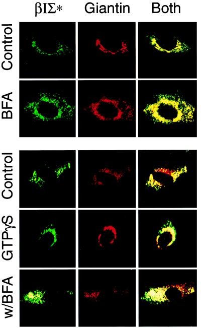
Figure 1.
Golgi βIΣ* spectrin contains PH domain epitopes and binds the Golgi complex in a GTPγS-dependent fashion. Four antispectrin antibodies were used to assess the distribution of the Golgi-associated spectrin. Two of these antibodies, MUS1 and MUS2, were specific for the PH domain of βIΣ2 spectrin. All gave similar distributions in NRK cells; the pattern of MUS1 staining is shown. This pattern largely overlaps that of giantin and is disrupted by BFA, and its reassembly is stimulated by GTPγS in permeabilized NRK cells. (Upper) Intact NRK cells were treated with 5 μg/ml of BFA for 5 min. (Lower) Streptolysin O-permeabilized NRK cells were treated with 50 μM GTPγS or with 1 μg/ml BFA for 5 min before and during permeabilization in the presence of 50 μM GTPγS (BFA). The immunofluorescence patterns shown are representative of the average pattern present in at least 80% of the cells for each treatment and observed in at least three experiments run in duplicate. Both represents the superimposition of the fluoresceine and Cy3 images, with yellow depicting the areas of overlap of the two antigens.

