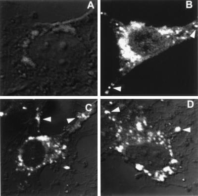
Figure 2.
Intracellular localization of ARD1 by immunofluorescence. NIH 3T3 cells were transfected with pcDNA3.1(ARD1) without a Kozak sequence (A), with pcDNA3.1(ARD1) with a Kozak sequence (B–D). Immunofluorescence images obtained after incubation of cells with anti-ARD1 (1:1,000) (A and B), anti-Nt-ARD1 (1:10,000) (C), and anti-Ct-ARD1 (1:10,000) (D) antibodies are shown. Arrows indicate labeled vesicular structures that are distant from the perinuclear area. Identical results were obtained with at least three different NIH 3T3 cell preparations and with COS 7 and HeLa cells.

