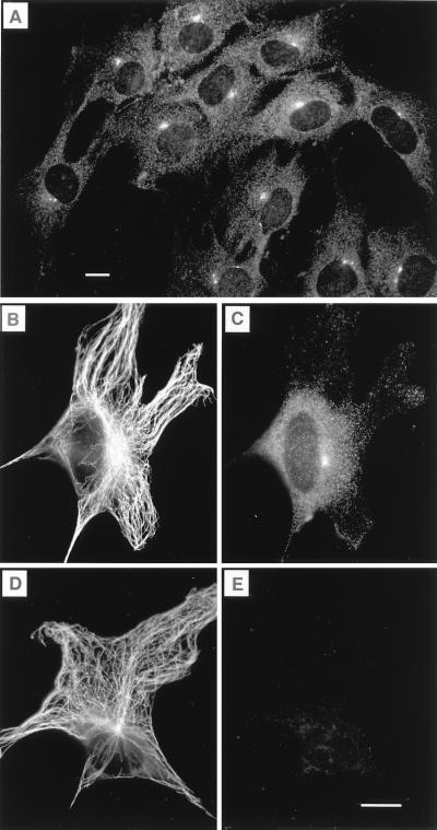
Figure 1.
Labeling of centrosomes with myosin-V antitail domain antibody in OC-k3 cells. (A) A field showing several interphase cells with intensely labeled foci in the perinuclear region. The three cells at the bottom appear to be in prophase (see Figs. 3 and 4). (B and C) A cell double-labeled with anti-β-tubulin and anti-myosin-V, respectively. The microtubule-organizing center coincided with the bright spot labeled with anti-myosin-V. (D and E) Preadsorption control cell labeled with anti-β-tubulin and anti-myosin-V plus 0.5 mg/ml chicken brain myosin-V, respectively. Preincubation with myosin-V did not affect the labeling by the anti-tubulin antibody, but completely blocked myosin-V labeling. Cells were fixed by using method ii (Materials and Methods). (Bars = 10 μm.)

