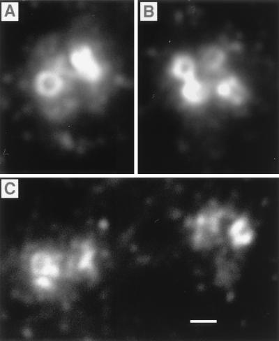
Figure 2.
High magnification of centrosomes labeled with the anti-tail antibody. (A) A pair of centrioles in an interphase cell. (B) Two pairs after replication. (C) Two pairs in early prophase just after poleward migration has begun. In each centrosome, the entire surface of the centrioles and a cluster of surrounding punctae were labeled. Cells were fixed by using method i (Materials and Methods). Centrosomes in A and B are from SV-k1 cells. Those in C are from B16-F10 cells. (Bar = 0.5 μm.)

