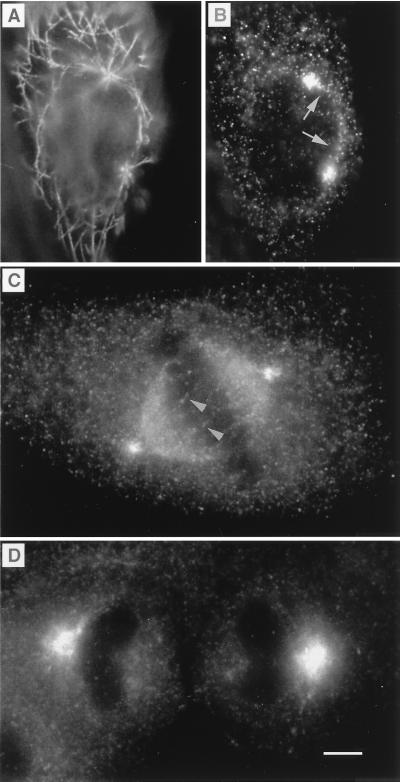
Figure 3.
Labeling of MDCK cells at different stages of the cell cycle. During prophase, the migrating microtubule-organizing centers were labeled with antitubulin (A), which coincided, at the poles, with myosin-V labeling (B). A trail of myosin-V labeling typically could be visualized running along the rim of the nucleus on the path of the migrating centrioles (arrow). At metaphase (C), myosin-V labeling could be visualized on spindle fibers and as a diffuse labeling throughout the spindle. To visualize detail in the regions of the poles and spindle, in metaphase cells (C) and in cells progressing from anaphase to telophase (D), brightness and contrast have been adjusted, so that the pronounced increase in diffuse cytoplasmic staining (text and Fig. 4) is not as obvious. Cells were fixed by using method i (Materials and Methods). Myosin-V antibodies were antitail (B), antihead, Hc (C), and antihead, Hf (D). (Bar = 5 μm.)

