Figure 4.
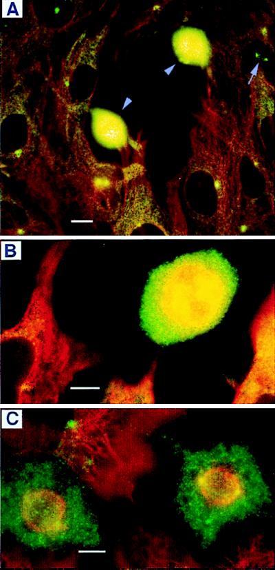
Double-labeling of cells with antibodies against myosin-V (green) and β-tubulin (red), showing the dramatic increase in the intensity of myosin-V staining in mitotic cells. The yellow color results from an overlap of the green and red. (A) Several stages of the cell cycle can be seen in SV-k1 cells, including interphase (most cells), prophase (arrow), and metaphase (intensely stained cells near the center). Higher magnification of metaphase cells (B, OC-k3 cells and C, SV-k1 cells) shows myosin-V labeling in the spindle and poles, and an intense, diffuse label throughout the cytoplasm. Cells were fixed by using method ii (Materials and Methods). Myosin-V antibodies were antitail (A and B) and antihead, Hf (C). (Bars = 10 μm.)

