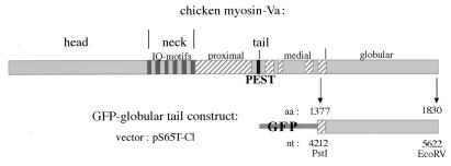
Figure 7.
Cloning strategy. Schematic diagram representing the linear domain structure of chicken myosin-Va and the GFP–globular tail fusion protein expressed from the construct obtained in the pS65T-C1 vector. The construct includes the full myosin-Va globular tail plus 45 aa residues from the medial tail as represented in the diagram, where the first and last amino acid and nucleotide (nt) positions are indicated.

