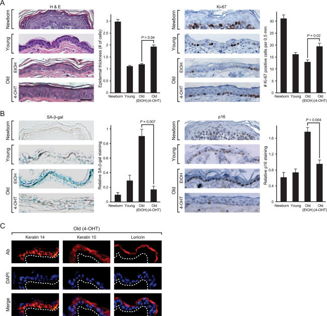
Figure 5.
Reversal of tissue characteristics of epidermal aging by NF-κB blockade. (A) NF-κB inhibition increases epidermal thickness and proliferation. (Left) Hematoxylin and eosin (H&E) staining of mouse epidermis. Average epidermal thickness (number of cell layers) is shown (±SEM). P-values show significant difference between old EtOH and old 4-OHT (Student’s t-test). Bar, 30 μm. (Right) Immunohistochemistry of Ki-67 protein expression in the epidermis of paraffin-embedded sections is shown (average number of Ki-67-positive cells ± SEM). (B) NF-κB blockade decreases senescence markers SA-β-gal and p16INK4A. (Left) SA-β-gal activity in the epidermis of frozen sections is shown (average SA-β-gal activity on a 0–3 scale ± SEM). (Right) Immunohistochemistry of p16INK4A protein in paraffin-embedded sections is shown (average p16INK4A staining on a 0–3 scale ± SEM). (C) Immunofluorescence of K14, K10, and loricrin (Ab, red) in frozen sections of old skin after NF-κB blockade. Nuclei are blue with DAPI counterstain. White dashed line indicates basement membrane.

