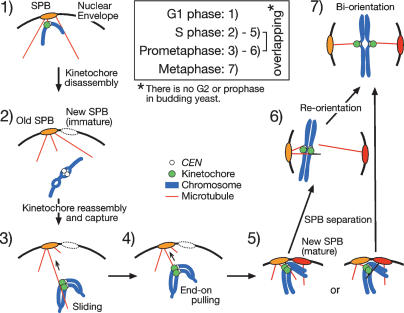
Figure 8.
Summary of kinetochore–microtubule interaction from G1 to metaphase in S. cerevisiae. (Step 1) Kinetochores are attached to microtubules (perhaps to the ends of microtubules; see Supplementary Note 6) in G1. (Step 2) Kinetochores are disassembled upon centromere DNA replication, and centromeres are detached from microtubules and move away from a spindle pole. (Step 3) Kinetochores are reassembled, captured by the lateral surface of microtubules, and transported poleward by sliding along the microtubule surface. (Step 4) Kinetochores are tethered at the ends of microtubules and often further transported poleward as microtubules shrink. (Step 5) Both sister kinetochores interact with microtubules from either the same or different SPBs. (Steps 6, 7) SPBs separate at the end of S phase, and reorientation of kinetochore–microtubule attachment leads to sister kinetochore biorientation.

