Figure 2.
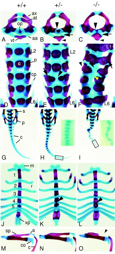
Skeletal structures of wild-type (A, D, G, J, and M), heterozygous (B, E, H, K, and N), and homozygous (C, F, I, L, and O) Pax1null newborn mice. (A–C) Superior view of atlas (at) and axis (ax). (A) Wild-type newborn. The odontoid process (op) of the axis is clearly separated from the arcus anterior atlantis (aa). vt, Ventral tubercle. (B) In heterozygous animals the ossified ventral tubercle is enlarged (small arrowhead) and is fused to the axis via cartilaginous bridges (arrow). Medial extensions of the pedicles of the axis are indicated by a large arrowhead. (C) In homozygous newborns the ossified ventral tubercle of the arcus anterior atlantis is enlarged (small arrowhead) and fused to the odontoid process of the axis (arrow). The pedicles of the axis extend medially to the ossification center of the vertebral body (large arrowhead). (D–F) Ventral view of lumbar vertebrae. (D) In wild-type newborns the ossification centers of the vertebral bodies (c) are clearly separated from the pedicles (p). Lumbar vertebrae L2 to L6 are shown, each separated by intervertebral discs (ivd). cp, Costal processes. (E) Lumbar vertebrae L2 to L6 of a heterozygous newborn with dual ossification center at L4 (arrowhead) are shown. The right part of the ossification center of L4 is fused to the right pedicle (arrow). (F) Lumbar vertebrae L1 to L6 of homozygous newborn. All lumbar vertebrae display fusions of the pedicles to the ossification centers (large arrow). In L3 to L5 vertebrae are saggitally split, ossification centers are missing (large arrowhead), and costal processes are lacking (small arrowhead). Intervertebral discs are reduced (small arrow), whereas in the sagittally split vertebrae a cartilaginous rod-like structure is formed medially (yellow arrowhead). Note that the malformations lead to skoliosis. (G–I) Dorsal views of sacrum and tail. (G) The sacral (s) and caudal (c) vertebrae of a wild-type newborn animal are shown; p, pelvic girdle. (H) In heterozygotes no difference to wild-type sacrum and tail can be observed. (Inset) A part of the tail (C16–19) in close-up. (I) Homozygous animals exhibit a strongly kinked tail. (Inset) The caudal segments C16–20 are shown in close-up. The vertebrae have a triangular shape caused by a drastically reduced posterior half of the segments. (J–L) Ventral view of the sternum. (J) In wild-type newborns the ossified sternebrae 1–5 are clearly separated by cartilaginous intersternebrae, to which the ribs attach. m, Manubrium; r, rib; xp, xiphoid process. (K) In heterozygotes the cartilaginous intersternebra between sternebrae 4 and 5 frequently exhibits a thin stripe of medio-sagittal ossification (arrowhead). (L) Homozygous newborns show complete ossification of the intersternebra connecting sternebrae 4 and 5 (arrowhead). (M–O) Left scapula in superior view. (M) Wild-type scapula connected to the clavicula (c) via the acromion (a); sp, spine of the scapula; co, coracoid process. (N) Scapula of heterozygote, indistinguishable from that of a wild-type newborn. (O) Scapula from homozygous newborn animal, displaying lack of a part of the ossified spine and of the cartilaginous acromion (arrowhead).

