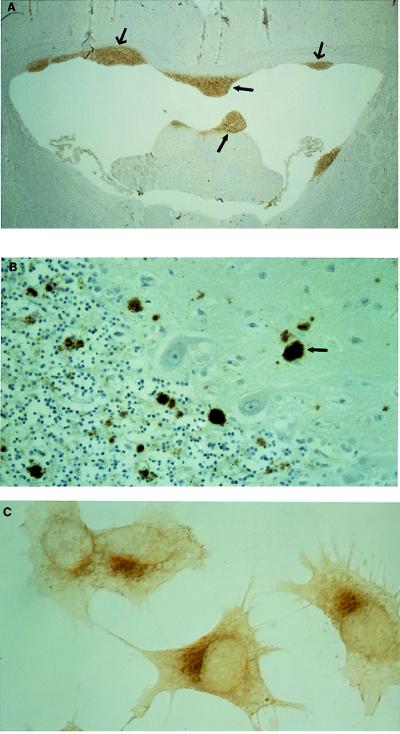
Figure 5.
Immunohistochemistry of PrPres in mouse brain, PrPres in CJD brain, and PrPc in a human neuroblastoma cell line. (A) Infected brain grafts from a transgenic mouse overexpressing PrPc, Tg20 mice, implanted in the ventricular wall of a Prnp−/− mouse. Paraffin sections were processed and stained with mAb 8H4. mAb 8H4 only stained the neurografts (arrows). (×20.) (B) Cerebellar tissues from the brain of a patient with CJD were processed and stained with mAb 8H4. Plaque-like PrP deposits (arrows) are diffusely distributed in the molecular and granular layers of the cerebellum. (× 150.) (C) A neuroblastoma cell line transfected with a construct expressing normal human PrPc was stained with 8H4. PrPc is distributed in the intracellular compartment with a Golgi-like distribution. (×430.)

