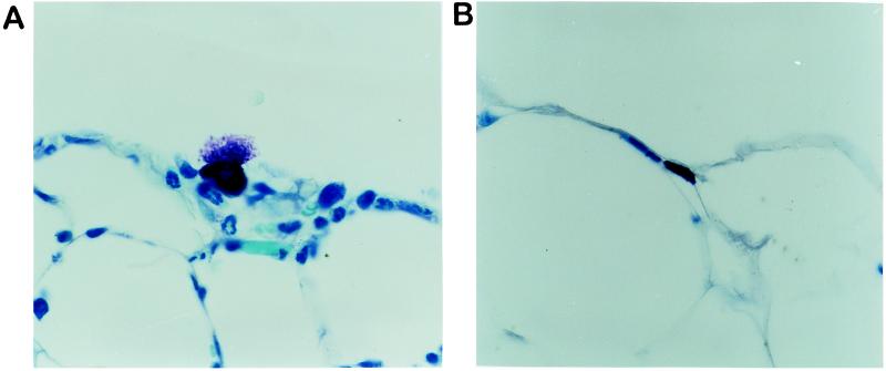
Figure 4.
Sections of omentum from an area immediately overlying a 16-hr PET i.p. implant (A) and from a distant site in the same animal that had no contact with the implant (B). These preparations were stained with toluidine blue, which permits the visualization of mast cell granules. Magnification, ×120.

