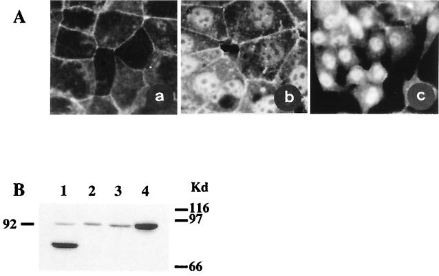
Figure 3.
β-catenin in human hepatoma cell lines. (A) β-catenin immunolocalization. Cultured human hepatoma cells were immunolabeled with rabbit polyclonal anti-β-catenin antibody. (a) PLC/PRF/5 hepatoma cells containing wild-type β-catenin and showing only membrane staining. Hepatoma cells with activated β-catenin showed nuclear staining. (b) Huh6 cells. (c) HepG2 cells. There was no labeling when the first anti-β-catenin antibody was omitted (not shown). (B) Western blot analysis. Truncated β-catenin in HepG2 cells (lane 1). Full-length β-catenin in PLC/PRF/5 and Hep3B cells (lanes 2 and 3). The G34V β-catenin mutation identified in the Huh6 cells led to accumulation of the protein (lane 4).

