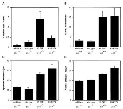
Figure 2.
Effect of p53 on apoptosis, proliferation, and hyperplasia in epidermis of K5 E2F1 transgenic mice. (A) Skin sections from line 1.0 transgenic and nontransgenic mice with and without wild-type p53 were subjected to TUNEL assays. Sections from four (nontransgenic) or five (transgenic) mice in each group were used to calculate the average number of apoptotic cells per 10 mm of interfollicular epidermis. (B) Line 1.0 transgenic and nontransgenic mice with and without p53 were injected with BrdUrd 30 min before sacrifice. At least 200 cells were counted to calculate the labeling index of interfollicular epidermis from two to four mice (2–13 weeks of age) in each group. (C) Skin samples were taken from line 1.0 transgenic and nontransgenic mice either with or without p53, ages 2–20 weeks. At least 10 measurements were taken from three to five different mice in each group to calculate the average thickness of the epidermis. (D) The same skin samples used in C were used to determine the average number of nuclei per 125 μm of epidermis.

