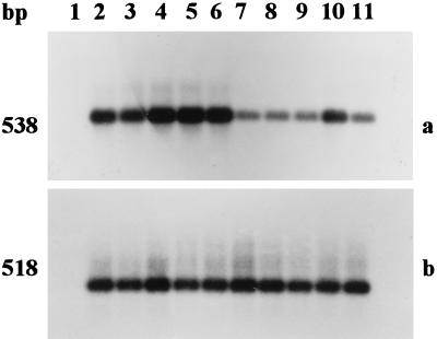
Figure 3.
Southern blot analysis of cDNA for human IGF-II (a) and β-actin (b) obtained after RT-PCR. The PCR products were separated by 1.8% agarose gel electrophoresis and transferred to Hybond N+ membranes. Hybridizations were performed with cDNA probes specific for hIGF-II or hβ-actin. The blots were washed under stringent conditions and exposed to autoradiography. Numbering is the same as in Fig. 2.

