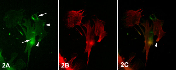
Figure 2.
Epifluorescent micrographs of epithelial cells of primary culture of normal breast tissue. (A) In the majority of the cells at the ventral surface, intense integrin alphavbeta3 immunofluorescence was observed at their perinuclear areas (arrows), and low diffused localization in the rest of the surface. Integrin alphavbeta3 aggregations (arrowheads) at the cell periphery were scarcely observed. (B) F-actin cytoskeleton, as visualized by rhodamine-conjucated phalloidin, was characterized by thin stress fibers arranged in parallel arrays across the cells. (C) Merged images of (A) and (B) showing the colocalization of stress fibers with integrin alphavbeta3 clusters at the sites of focal contacts (arrowhead). [750×].

