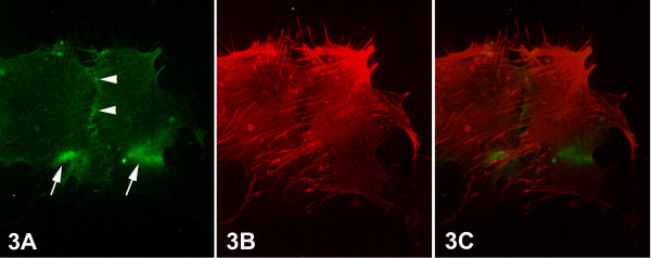
Figure 3.
Epifluorescent micrographs of epithelial cells of primary culture of breast cancer tissue. (Figs. 3A) Bright aggregations of integrin alphavbeta3 immunofluorescence – indicating integrin clustering – were observed at the marginal areas of the ventral surface of the cells (arrows). Fine granular integrin alphavbeta3 immunofluorescence was also observed at the boundary between the cells in contact (arrowheads). (Fig. 3B) F-actin cytoskeleton, as visualized by rhodamine-conjucated phalloidin, appeared well developed, constituted by numerous thick stress fibers, which were arranged towards the integrin alphavbeta3 immunolocalizations, as demonstrated also in the merged images (Fig. 3C). [750×].

