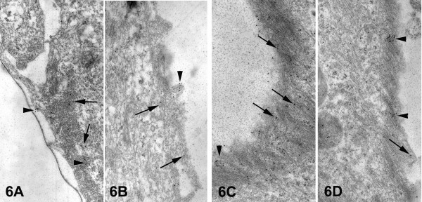
Figure 6.
Electron micrographs showing the integrin alphavbeta3 immunogold labeling in epithelial cells of primary cultures of normal breast tissues (A, B) and of breast cancer biopsies (C, D). In both cases staining pattern is essentially the same i.e. mostly single gold particles (arrows), and occasionally aggregates (arrowheads) over the region of the stress fibers. However, in cancer epithelial cells an increased integrin alphavbeta3 immunogold labeling is observed. [25,250×].

