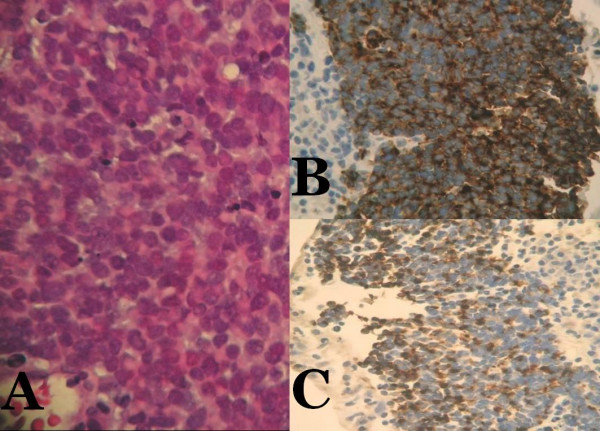
Figure 4.

Microscopic examination reveals tumor composed of monotonous round cells showing scant eosinophilic cytoplasmic rim, round and vesicular nuclei with finely granular and dusty chromatin and multiple nucleoli (A, hematoxylin and eosin ×100). Tumor cells are positive for synaptophysin (B, ×100) and CK 20 (C, ×100).
