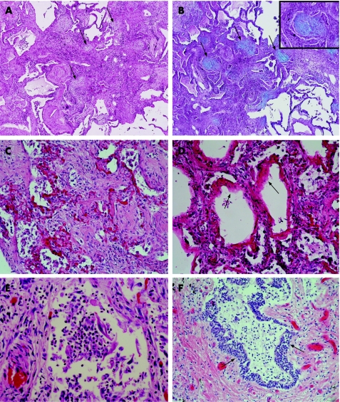
Figure 1 (A) Fibroblast foci (arrows) in usual interstitial pneumonia (UIP) stained with haematoxylin‐eosin (HE); original magnification ×25.5. (B) Fibroblast foci (arrows) in UIP stained with Alcian blue‐periodic acid Schiff (AB‐PAS); original magnification ×25.5. Inset: a fibroblast focus at higher magnification; original magnification ×2.8. (C) Diffuse alveolar damage (DAD) in a necroscopic lung sample from a patient with idiopathic UIP; original magnification ×63.8. (D) Hyaline membranes (arrows) of DAD in a necroscopic sample from a patient with idiopathic UIP; original magnification ×63.8. (E) Intra‐alveolar neutrophils in a necroscopic sample from a patient with idiopathic UIP; original magnification ×255. (F) Metaplastic squamous type alveolar epithelium (arrows) in a necroscopic sample from a patient with idiopathic UIP; original magnification ×63.8.

An official website of the United States government
Here's how you know
Official websites use .gov
A
.gov website belongs to an official
government organization in the United States.
Secure .gov websites use HTTPS
A lock (
) or https:// means you've safely
connected to the .gov website. Share sensitive
information only on official, secure websites.
