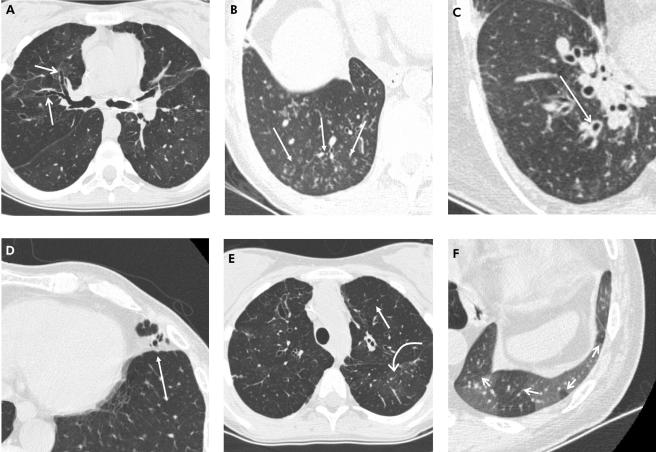
Figure 1 Representative CT images showing the CTBO scoring system abnormalities. A collection of transaxial 1.25 mm CT sections in different patients viewed at lung window and level setting (width 1500 HU, level −500 HU) showing (A) bronchiectasis (arrows) identified by the absence of normal bronchial diameter tapering; (B) peripheral mucus plugging shown as multiple centrilobular nodules (arrows); (C) dilated bronchus with associated wall thickening (arrow); (D) lingular consolidation (arrow) shown as an area of increased density obscuring the underlying pulmonary vasculature; (E) generalised mosaic pattern in both upper lobes shown by areas of decreased attenuation and vessel size (straight arrow) compared with regions of normal attenuation and normal vessel size (curved arrow); and (F) expiratory image showing areas of air trapping (arrows).

An official website of the United States government
Here's how you know
Official websites use .gov
A
.gov website belongs to an official
government organization in the United States.
Secure .gov websites use HTTPS
A lock (
) or https:// means you've safely
connected to the .gov website. Share sensitive
information only on official, secure websites.
