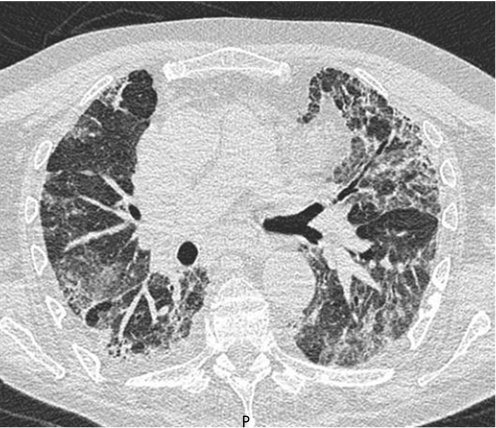
Figure 3 Typical CT scan from a patient with non‐specific interstitial pneumonia. Note the less peripheral distribution of disease than in idiopathic pulmonary fibrosis and the widespread ground‐glass change, but with clear evidence of traction of the airways indicating that at least some, but not necessarily all, of this ground change is due to fine fibrosis.

An official website of the United States government
Here's how you know
Official websites use .gov
A
.gov website belongs to an official
government organization in the United States.
Secure .gov websites use HTTPS
A lock (
) or https:// means you've safely
connected to the .gov website. Share sensitive
information only on official, secure websites.
