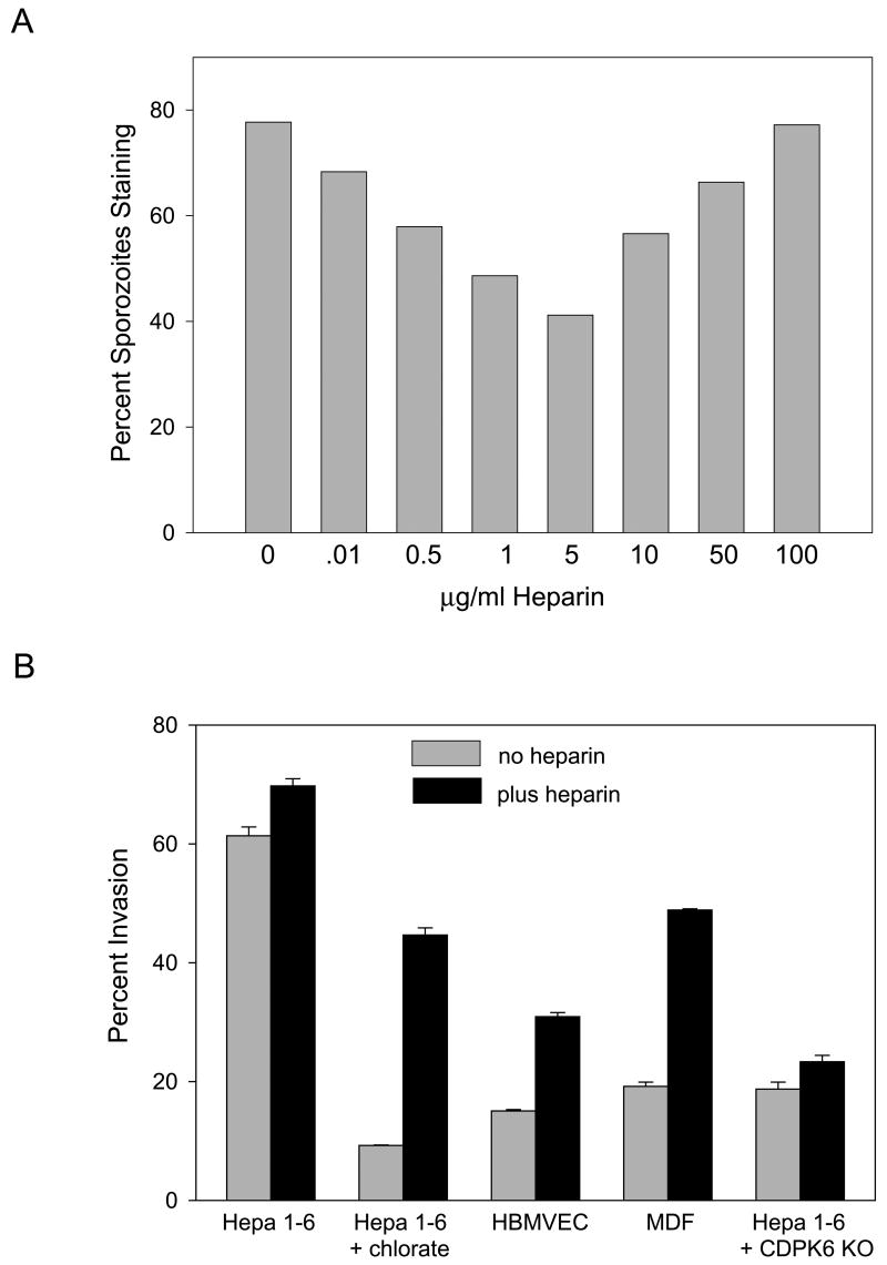
Figure 4. Soluble heparin induces CSP cleavage and enhances sporozoite invasion of nonpermissive cells.
(A) CSP cleavage after a 5 min incubation with the indicated concentrations of heparin was quantified by fixing and staining sporozoites with antisera specific for full-length CSP. 200 sporozoites per well were counted and shown is the percentage of sporozoites staining. This experiment was repeated 3 times with identical results. (B) Sporozoite invasion of the indicated cell line was quantified by adding wild type or CDPK-6 mutant (CDPK6 KO, see below) sporozoites preincubated for 5 min with medium alone (grey bars) or medium containing 5 μg/ml heparin (black bars) to Hepa 1–6 cells grown in the absence or presence of chlorate, HBMVEC or MDF cells. After 1 hr, cells were fixed, stained and the numbers of intracellular and extracellular sporozoites were counted. Shown are means ± SD of triplicates. This experiment was repeated twice with identical results.

