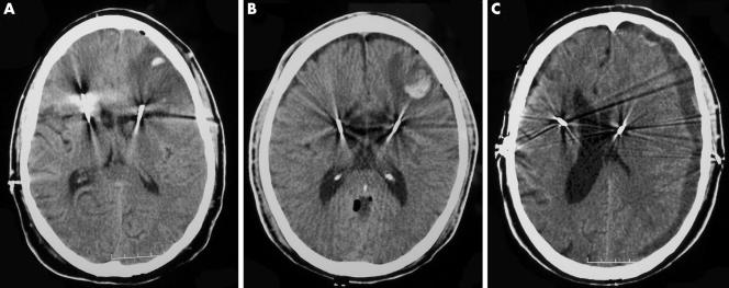
Figure 1 Axial CT images demonstrating the three haemorrhagic related morbidities in our 100 patient series. (A) Low intensity left frontal lobe lesion consistent with venous infarction (this patient had a history of anti‐factor XI antibodies); (B) lesion consistent with small frontal lobe haemorrhage; (C) hypointense left subdural collection consistent with subacute haematoma with significant midline shift. Each patient recovered without permanent neurological sequelae.

An official website of the United States government
Here's how you know
Official websites use .gov
A
.gov website belongs to an official
government organization in the United States.
Secure .gov websites use HTTPS
A lock (
) or https:// means you've safely
connected to the .gov website. Share sensitive
information only on official, secure websites.
