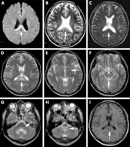
Figure 1 Axial diffusion weighted image from case 1 demonstrates an increased signal intensity area in the splenium of the corpus callosum (A). Axial T2 weighted magnetic resonance image demonstrates hyperintense lesions in the same region (B). Another T2 weighted image obtained 1 month later discloses near resolution of the lesions (C). T2 weighted axial image in case 2 shows bilateral symmetric high signal intensities in the splenium of the corpus callosum (D), the globus pallidus (E), the periaqueductal grey matter of the midbrain (F), the pontine tegmentum (G), the dentate nucleus (G), and the medulla oblongata (H). These lesions were hypointense on T1 weighted images (I).

An official website of the United States government
Here's how you know
Official websites use .gov
A
.gov website belongs to an official
government organization in the United States.
Secure .gov websites use HTTPS
A lock (
) or https:// means you've safely
connected to the .gov website. Share sensitive
information only on official, secure websites.
