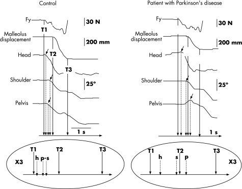
Figure 2 Single trial recording of body rotation: left, control; right, a patient with Parkinson's disease. Top: kinetic (Fy: horizontal forces in the sagittal plane), kinematic (malleolus displacement) and angular data. The horizontal rotation around the vertical axis (yaw), angular variation of the head, the shoulders and the pelvis are shown. The small arrows indicate the onset of rotation of each segment. Bottom: representation of the temporal organisation of the rotation. Controls begin shoulder and pelvis rotation simultaneously with their axial body rotation, whereas patients with Parkinson's disease start shoulder (s) rotation before pelvis (p) rotation. Trunk rotation is uncoupled. T1, start of the postural phase (first variation in the horizontal forces in the sagittal plane); T2, end of the postural phase corresponding to the beginning of the movement phase (first variation in the velocity of the malleolus marker); T3, end of the movement phase (first contact of the foot with the floor).

An official website of the United States government
Here's how you know
Official websites use .gov
A
.gov website belongs to an official
government organization in the United States.
Secure .gov websites use HTTPS
A lock (
) or https:// means you've safely
connected to the .gov website. Share sensitive
information only on official, secure websites.
