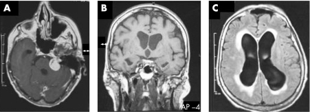
Figure 1 (A) T1 weighted MRI scan demonstrated a tumour in the left cerebellopontine angle, compatible with a vestibular schwannoma which was accompanied by enlargement of the ventricles adjacent to the caudate nucleus. The aqueduct is patent (B). Fluid attenuated inversion recovery series showed periventricular effusion (C).

An official website of the United States government
Here's how you know
Official websites use .gov
A
.gov website belongs to an official
government organization in the United States.
Secure .gov websites use HTTPS
A lock (
) or https:// means you've safely
connected to the .gov website. Share sensitive
information only on official, secure websites.
