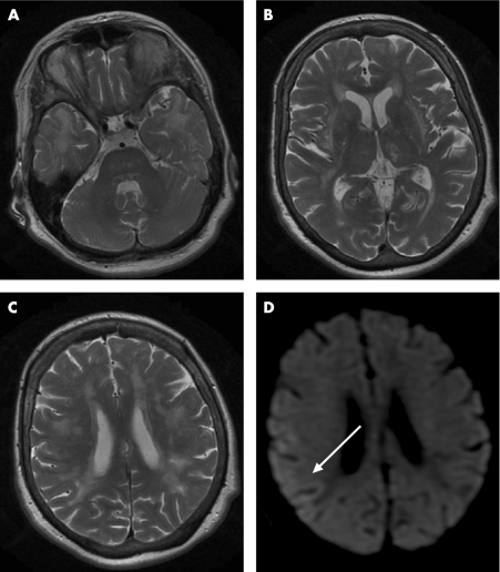
Figure 1 T2 weighted MRI scans showing diffuse hyperintense signal abnormalities with prominent involvement of both temporal lobes (A), both thalami, external and extreme capsules (B), as well as widespread areas of the white matter (C). Diffusion weighted imaging does not show evidence of an acute ischaemic strokes but demonstrates a weak and diffuse signal alteration in the right parieto‐occipital cortex 24 h before the first EEG (arrow; D).

An official website of the United States government
Here's how you know
Official websites use .gov
A
.gov website belongs to an official
government organization in the United States.
Secure .gov websites use HTTPS
A lock (
) or https:// means you've safely
connected to the .gov website. Share sensitive
information only on official, secure websites.
