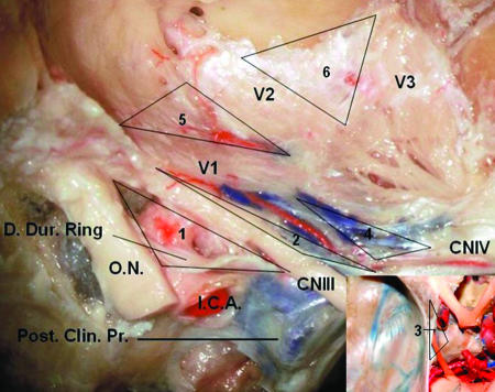
Figure 1.
View of the right cavernous sinus with triangle identification. The anterior clinoid process was removed, exposing the clinoidal triangle (1). The outer layer of the dura has been peeled away, exposing the supratrochlear (2), infratrochlear (4), anteromedial middle fossa (5), and anterolateral middle fossa (6) triangles. The oculomotor triangle (3) is delimited in the smaller picture. The cranial nerve (CN) IV is running parallel to the tentorial artery. D. Dur., distal dural; O.N., optic nerve; I.C.A., internal carotid artery; Post. Clin. Pr., posterior clinoid process.

