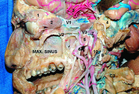
Figure 6.
Lateral view of the CS and adjacent regions. The lateral and anterior walls of the maxillary sinus were removed. The walls of the orbit and orbital fat were removed. The temporal lobe is displaced posteriorly to expose the CS. The dura of the middle fossa was peeled away and the bone of the middle fossa floor was drilled out to show the anatomical relationship between the temporal and infratemporal fossa structures. Mastoidectomy was performed, preserving the mastoid tip. 1, lacrimal gland; 2, rectus lateralis muscle; 3, middle cerebral artery; 4, otic capsule (semicircular canals); 5, Eustachian tube; 6, middle meningeal artery; 7, internal carotid artery (intrapetrous portion); 8, facial nerve; 9, vertebral artery; 10, styloid process. CS, cavernous sinus; Max., maxillary.

