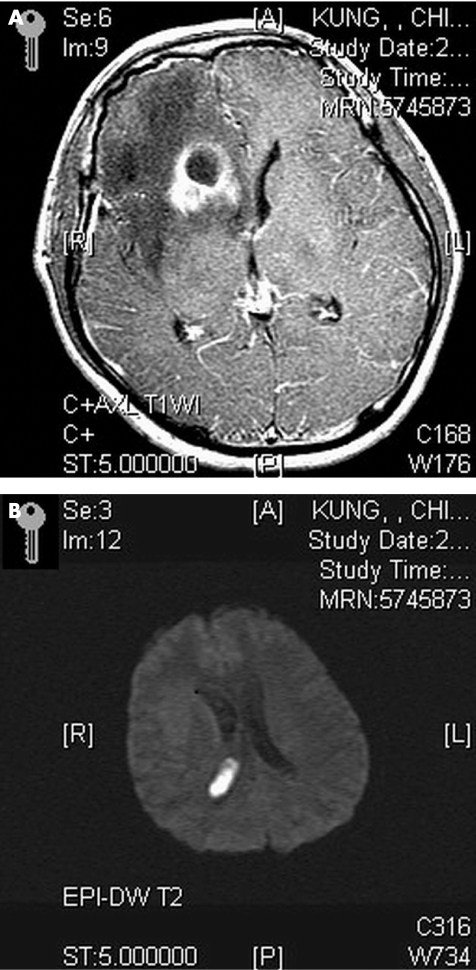
Figure 1 (A) Magnetic resonance imaging axial view T1‐weighted image with gadolinium‐diethylenetriaminepentaacetic acid administration shows a ring‐enhancing lesion in the right frontal horn, and increased enhancement in the leptomeninges and periventricular region. (B) Diffusion‐weighted image shows a hyperintense lesion in the right lateral ventricle consistent with intraventricular debris.

An official website of the United States government
Here's how you know
Official websites use .gov
A
.gov website belongs to an official
government organization in the United States.
Secure .gov websites use HTTPS
A lock (
) or https:// means you've safely
connected to the .gov website. Share sensitive
information only on official, secure websites.
