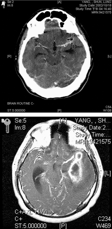
Figure 3 A 34‐year‐old man presented with postneurosurgical central nervous system infection, with an initial Glasgow Coma Score of E3V4M6. (A) Computed tomography scan of the brain with intravenous contrast administration showing a small ring‐enhanced lesion in the left Sylvian fissure region. (B) Magnetic resonance imaging axial view T1‐weighted image with gadolinium administration performed 1 month later showing multiloculated enhanced lesions in the left temporal region touching the ventricular wall of the temporal horn.

An official website of the United States government
Here's how you know
Official websites use .gov
A
.gov website belongs to an official
government organization in the United States.
Secure .gov websites use HTTPS
A lock (
) or https:// means you've safely
connected to the .gov website. Share sensitive
information only on official, secure websites.
