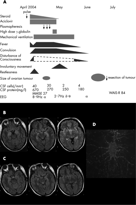
Figure 1 Clinical course of the patient, magnetic resonance image of the brain and immunolabelling of live rat hippocampal neurones with the patient's cerebrospinal fluid (CSF). (A) Clinical course of the patient. The symptoms and laboratory data were improved before the tumour resection. (B) MRI fluid‐attenuated inversion recovery images of the brain in April 2004 showed areas of hyperintensity in the medial temporal lobes, cingulate gyrus, insular regions and hippocampus. (C) These abnormalities had resolved by June 2004. (D) The patient's antibodies, which colocalised with EFA6A, showed intense immunolabelling of the neuronal cell membranes and processes, using methods previously reported2. WAIS‐R, Weschler Adult Intelligence Scale—Revised.

An official website of the United States government
Here's how you know
Official websites use .gov
A
.gov website belongs to an official
government organization in the United States.
Secure .gov websites use HTTPS
A lock (
) or https:// means you've safely
connected to the .gov website. Share sensitive
information only on official, secure websites.
