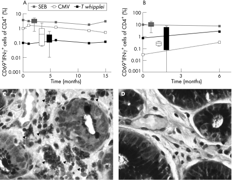
Figure 1 Stimulation of whole blood (A) and duodenal lymphocytes (B) with staphylococcus enterotoxin B (SEB 2 μg/ml), cytomegalovirus (CMV 1.2 μg/ml) and Tropheryma whipplei lysate (T whipplei 107 bacteria/ml) (for methods see Working Group on Coeliac Disease of the United European Gastroenterology Week in Amsterdam5). The curves represent the time course of per cent CD69+ interferon γ+ (IFNγ) cells of CD4+ lymphocytes of the patient, and box and whisker values of unaffected controls (10 subjects during follow‐up of ulcer disease without Whipple disease served as controls). (C, D) Periodic acid–Schiff (PAS) staining of biopsies from the antrum. (C) T whipplei infected type I macrophages (arrowheads, see also von Herbay and colleagues9) in the antrum before the onset of therapy. (D) PAS negative antrum 18 months after the onset of therapy.

An official website of the United States government
Here's how you know
Official websites use .gov
A
.gov website belongs to an official
government organization in the United States.
Secure .gov websites use HTTPS
A lock (
) or https:// means you've safely
connected to the .gov website. Share sensitive
information only on official, secure websites.
