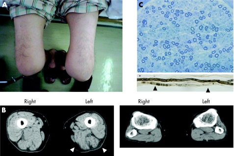
Figure 1 Clinical features and biopsy results. (A) The patient's lower extremities. The left thigh is visibly atrophic and exhibits redness in its entirety. (B) Muscle atrophy of the thigh (left panel) was confirmed by computed tomography (arrowheads). There was no clear muscle atrophy in the lower legs (right panel). (C) Transverse section of sural nerve specimen stained with toluidine blue; there was a slight decrease in myelinated fibres. Teased fibres showed axonal degeneration (arrowheads). Informed consent was obtained for publication of this figure.

An official website of the United States government
Here's how you know
Official websites use .gov
A
.gov website belongs to an official
government organization in the United States.
Secure .gov websites use HTTPS
A lock (
) or https:// means you've safely
connected to the .gov website. Share sensitive
information only on official, secure websites.
