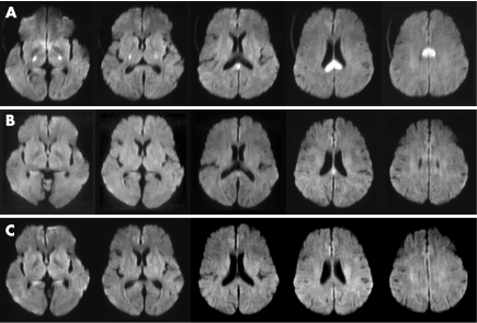
Figure 1 (A) Diffusion‐weighted imaging (DWI) on admission showing hyperintense lesions in the splenium of the corpus callosum and the bilateral posterior limbs of the internal capsules. (B) DWI obtained 2 h after glucose infusion showing almost full recovery except for a small part of the splenium of the corpus callosum. (C) DWI obtained 2 days after glucose infusion showing complete regression of the hyperintense lesions.

An official website of the United States government
Here's how you know
Official websites use .gov
A
.gov website belongs to an official
government organization in the United States.
Secure .gov websites use HTTPS
A lock (
) or https:// means you've safely
connected to the .gov website. Share sensitive
information only on official, secure websites.
