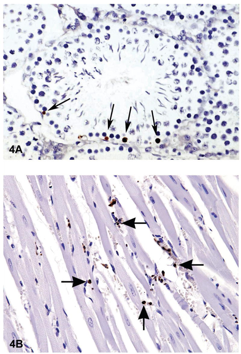
Figure 4.

There are many ways of detecting apoptosis at different stages on histological sections. One commonly used method is called TUNEL (Terminal deoxynucleotidyl Transferase Biotin-dUTP Nick End Labeling). One of the major characteristics of apoptosis is the degradation of DNA after the activation of Ca/Mg dependent endonucleases. This DNA cleavage leads to strand breaks within the DNA. However, necrosis can also result in similar DNA cleavage. Therefore an additional method should be used to confirm apoptosis. The TUNEL method identifies cells with DNA strand breaks in situ by using terminal deoxynucleotidyl transferase (TdT) to transfer biotin-dUTP to the cleaved ends of the DNA. The biotin-labeled cleavage sites are then detected by reaction with HRP (horse radish peroxidase) conjugated streptavidin and visualized by DAB (diaminobenzidine), which gives a brown color. The tissue is typically counterstained with Toluidine blue to allow evaluation of tissue architecture. Figure 4A a photomicrograph of a section of testes from a B6C3F1 mouse that was in a National Toxicology Program bioassay. The TUNEL assay was used on this section of tissue to detect apoptotic cells. The seminiferous tubule is cut in cross section and has sperm, spermatogonia and spermatocytes in various stages of development. The arrows point to labeled (dark brown) apoptotic spermatogonia in the basal layer of the seminiferous epithelium. Figure 4B is an image of myocardium from 14 week-old rat treated with ephedrine (25 mg/kg) and caffeine (30 mg/kg). There are multiple apoptotic cells that have stained brown with the TUNEL method (arrows).
