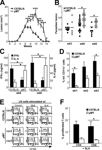
Figure 7.
Increased lesion progression in μMT mice due to decreased numbers of L. major–infected lesional DC and impaired T cell priming. Groups of ≥5 C57BL/6 or B cell–deficient μMT mice were infected into ear skin with 103 metacyclic promastigotes. (A) Lesion development was monitored over the course of >3 mo (mean ± SEM, *, P ≤ 0.05, **, P ≤ 0.005, and ***, P ≤ 0.002, n ≥ 3). (B) Lesional parasite loads were determined; bars indicate arithmetic means. Pooled data of two to three experiments are shown. (C) LN cells were harvested and antigen-specific cytokine release was determined after 48 h using ELISA specific for murine IFN-γ and IL-4 (mean ± SEM, n ≥ 5, *, P ≤ 0.05). (D) Inflammatory ear cells were isolated and CD11c+ DCs were purified using flow sorting. The percentage of infected DCs was determined on cytospins (mean ± SEM, n = 3, *, P ≤ 0.05). (E and F) Antigen-specific proliferation of CD4+ and CD8+ T cells was determined in week 6 after infections. LN cells were labeled with CFSE and subcultured in the presence of soluble Leishmania antigen (SLA). T cells were pregated using staining for CD4 or CD8. For each mouse, the relative number of Leishmania-reactive cells (in percentage of total CD4+ or CD8+ T cells) was calculated in antigen-stimulated compared with untreated control cultures (mean ± SEM, n = 2, *, P ≤ 0.05).

