Figure 2.
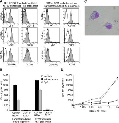
IPCs and DCs developed in culture from huFlt3-transduced Flt3− progenitors are functional. (A) Contour plots indicate surface markers (closed histogram) and respective isotype controls (open histogram) on CD11c+B220+ and CD11c+B220− cells derived from huFlt3L-Ig– and SCF-cultured huFlt3-transduced Flt3− progenitors. CD11c+B220+ and CD11c+B220− cells show typical phenotypes of IPCs and DCs, respectively. Results are from one representative experiment out of three. (B) Sorted CD11c+B220+ IPCs but not CD11c+B220− DCs, both derived from either huFlt3-transduced Flt3− progenitors or GFP-transduced Flt3+ progenitors, produce IFN-α upon influenza virus or CpG stimulation. Culture supernatants were collected after 24 h and analyzed by ELISA. Results are from one representative experiment out of three. (C) About half of day 8 progeny from huFlt3-transduced Flt3− progenitors display typical DC morphology. Giemsa-stained cytospin, photographed at a magnification of 40. (D) In vitro–generated CD11c+B220− cells from retrovirus-transduced progenitors are efficient stimulators of allogeneic T cells. Graph depicts thymidine incorporation of 2 × 10⁵ allogeneic BALB/c spleen CD4+ T cells incubated with graded numbers (x axis) of sorted CD11c+B220− DCs derived from huFlt3-transduced Flt3− progenitors (•), CD11c+B220+ IPCs derived from huFlt3-transduced Flt3− progenitors (▪), or CD11c+B220− DCs derived from GFP-transduced Flt3+ progenitors (△), and CD11c+B220+ IPCs derived from GFP-transduced Flt3+ progenitors (⋄). Results are from one representative experiment out of three.

