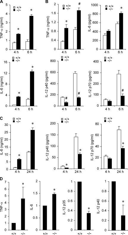
Figure 3.
Knockout of Mkp-1 alters cytokine expression in macrophages. (A) Cytokine production by IFN-γ–primed resident peritoneal macrophages. Resident peritoneal macrophages primed with IFN-γ overnight were stimulated with LPS for 4 and 6 h. Cytokine concentrations in the medium were analyzed by ELISA. Data are presented as the mean ± SEM (n = 3 in each group). *, Mkp-1 −/− different from Mkp-1 +/+, P < 0.001. (B) Cytokine production by thioglycollate-elicited peritoneal macrophages after stimulation with 100 ng/ml LPS for 4 and 6 h. Data are presented as mean ± SEM (n = 4 in each group). *, Mkp-1 −/− different from Mkp-1 +/+, P < 0.05; #, Mkp-1 −/− different from Mkp-1 +/+, P < 0.005. (C) Cytokine production by unprimed resident peritoneal macrophages from both Mkp-1 +/+ and Mkp-1 −/− mice. Resident peritoneal macrophages were stimulated with 100 ng/ml LPS for the indicated times, and cytokine concentrations in the medium were assayed by ELISA. Data are presented as the mean ± SEM (n = 6 in each group). *, Mkp-1 −/− different from Mkp-1 +/+, P < 0.001. (D) Expression of TNF-α, IL-6, IL-12p35, and IL-12p40 mRNA in LPS-stimulated wild-type and Mkp-1 −/− resident peritoneal macrophages. Resident peritoneal macrophages were primed overnight with 50 U/ml IFN-γ and then stimulated with 100 ng/ml LPS for 4 h. qRT-PCR was performed to assess the levels of mRNA for TNF-α, IL-6, IL-12p35, and IL-12p40. The relative mRNA levels were normalized to the GAPDH mRNA. Values represent expression levels relative to those in wild-type cells. Data are means ± SEM of three independent experiments. *, different from Mkp-1 −/− cells, P < 0.05.

