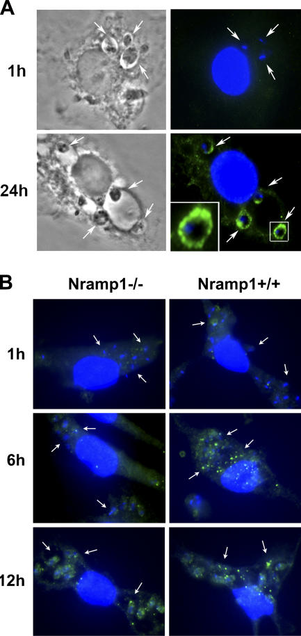
Figure 2.
L. amazonensis LIT1 is expressed by amastigotes residing intracellularly, and expression is accelerated in Nramp1+/+ macrophages. Mouse BMmø were infected with L. amazonensis axenic amastigotes for 1 h, washed, and further incubated for the indicated periods of time, followed by fixation, permeabilization, and immunofluorescence with antibodies specific for LIT1. (A) C57BL/6 BMmø (Nramp1−/−). Phase contrast (left) and fluorescence microscopy (right) images are shown. LIT1 was only detected 24 h after infection in a pattern consistent with localization on the plasma membrane of amastigotes (the inset shows an enlarged image). (B) C57BL/10ScSn (Nramp1−/−) or B10.L-Lsh (Nramp1+/+) BMmø. Punctate LIT1 immunofluorescence was detected after 12 h in the Nramp1−/− BMmø and after 6 h in the Nramp1−/− BMmø. Antibodies to LIT1 are labeled in green, and the host cell and parasite's DNA are stained in blue (DAPI). Arrows in A and B point to intracellular parasites. The images were acquired and enhanced for contrast under identical conditions.

