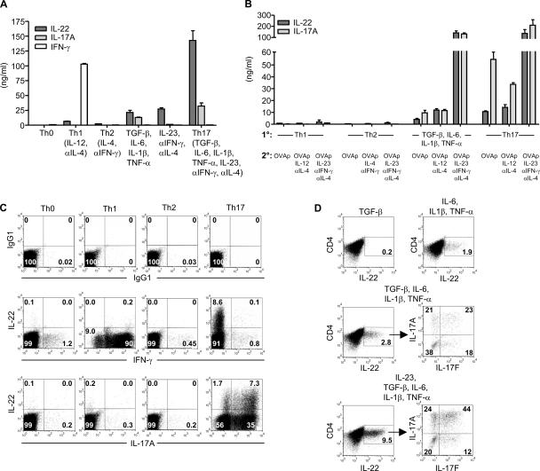
Figure 2.
Th17 cells express IL-22 protein. (A) Naive DO11 T cells were activated with irradiated splenocytes, OVAp, and various cytokines and antibodies. IL-22, IL-17A, and IFN-γ concentrations were measured on day 5. (B) Naive DO11 cells were differentiated under Th1, Th2, or Th17 conditions, or with TGF-β, IL-6, IL-1β, and TNF-α for 7 d. After resting overnight, cells were then restimulated with OVAp, IL-2, irradiated splenocytes, and combinations of cytokines and antibodies. IL-22 and IL-17A concentrations were measured on day 5. (C) Intracellular cytokine staining was performed on cells from A on day 5. Plots are gated on KJ126+CD4+ cells. (D) Naive DO11 T cells were activated as in A with the indicated cytokines. Intracellular cytokine staining was performed on day 5. Data shown are representative of three independent experiments. Error bars are SD.

