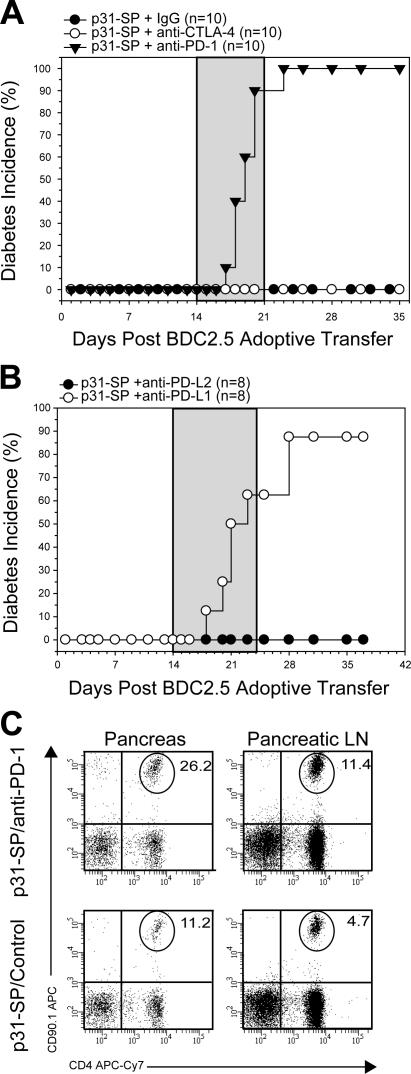
Figure 4.
PD-1 and PD-L1, but not CTLA-4, maintain peripheral tolerance. (A) p31-SP–tolerized NOD mice that received BDC T cells were treated with anti–PD-1, anti–CTLA-4, or IgG control (days 14, 16, 18, 20, 22, and 24; gray shaded area) to determine the role of these inhibitory molecules during tolerance maintenance. Diabetes incidence from p31-SP anti–PD-1 (100%; n = 10; ▾), p31-SP anti–CTLA-4 (0%; n = 10; ◯), and p31-SP IgG (0%; n = 10; •) is shown. (B) p31-SP–tolerized mice were injected with anti–PD-L1 or anti–PD-L2 on days +14, 16, 18, 20, 22, and 24 (gray shaded area) to determine which PD-1 ligands were important for tolerance. Diabetes incidence from p31-SP anti–PD-L1 (87.5%; n = 8; ◯) and complete protection in p31-SP anti–PD-L2 (0%; n = 8; •) is shown. (C) Diabetogenic CD4+CD90.1+ BDC2.5 T cell percentages from the pancreas and pLN were determined 7 d after anti–PD-1 treatment, illustrating an increase after anti–PD-1 injection. Results shown are representative from at least two independent experiments.

