Figure 2.
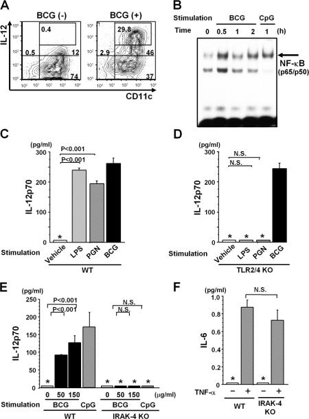
Activation of DCs by BCG. IL-12 production (A) and NF-κB activation (B). (A) Intracellular staining of BM-DCs with anti–IL-12p40/p70 and anti-CD11c mAbs with or without in vitro BCG (50 μg/ml) treatment for 12 h. BCG-treated BM-DCs (10,000 cells) were analyzed by FACS, and the number in each panel indicates the percentage of total cells. (B) NF-κB activation. 2 × 10⁵ BM-DCs were stimulated with or without 50 μg/ml BCG or 1 μM CpG in vitro. NF-κB activity was determined by EMSA. (C and D) No requirement of TLR2 and TLR4 in BCG-mediated IL-12 production. 2 × 10⁵ BM-DCs derived from WT (C) or TLR2/4 double KO (D) mice were stimulated in vitro with or without 10 μg/ml LPS, 10 μg/ml PGN, or 150 μg/ml BCG for 48 h, and IL-12p70 levels were measured by ELISA. (E and F) Requirement of IRAK-4 for IL-12 production. 2 × 10⁵ BM-DCs were assayed for IL-12p70 by ELISA after stimulation with 0, 50, or 150 μg/ml BCG or 1 μM CpG (E), and for IL-6 with 10 ng/ml TNF-α stimulation for 48 h (F). In C–F, values are expressed as mean ± SD of triplicate cultures. The asterisks (*) indicate that the levels were below the detection limits for IL-12p70 (<62.5 pg/ml) and IL-6 (<15.6 pg/ml). N.S., not significant. All experiments were repeated twice with similar results.

