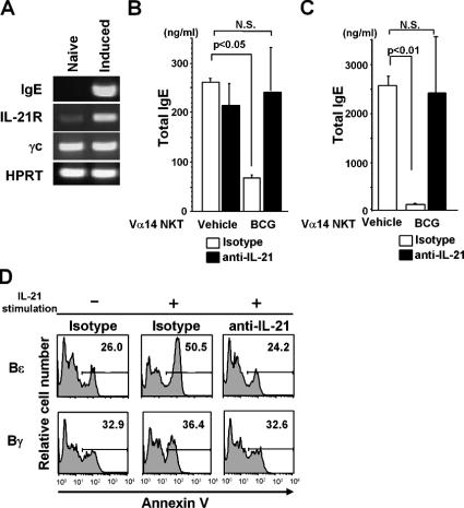
Figure 4.
IL-21–mediated Bɛ cell apoptosis. (A) RT-PCR analysis. Expression of IgE (Cɛ), IL-21R, and γc was investigated in naive B (left) and Bɛ (right) cells. (B) Suppression of IgE production in naive B cell cultures. Naive B cells and Vα14 NKT cells (105 each) were cocultured in the presence of sCD40L and IL-4. (C) Suppression of IgE production in the Bɛ cell culture. 105 Vα14 NKT cells were added to the Bɛ cell (105) cultures. In B and C, 20 μg/ml anti–IL-21 mAb or isotype control mAb was added at the same time as the Vα14 NKT cells. The concentration of total IgE was measured by ELISA in triplicate. Values are expressed as mean ± SD. N.S., not significant. The experiments were repeated three times with similar results. (D) IL-21–mediated Bɛ cell apoptosis. 2 × 10⁵ Bɛ and Bγ cells were generated and then further cultured with or without 30 ng/ml IL-21 for 30 h. Annexin V staining was then performed. The numbers represent percentage of the gated cells. Annexin V+ cells among Bɛ and Bγ cells just before IL-21 treatment was 25.7 and 29.2%, respectively (not depicted). The experiments were repeated three times with similar results.

