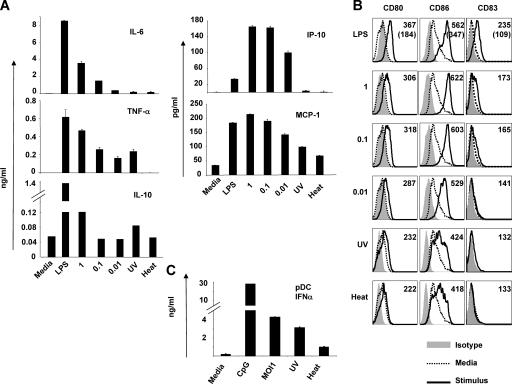
Figure 1.
YF-17D activates human monocyte–derived DCs and plasmacytoid DCs. Human mDCs were cultured with LPS; YF-17D at an MOI of 1, 0.1, or 0.01; or they were YF-17D UV or heat inactivated. Cytokines (A) and costimulatory molecule expression (B) were measured after 48 h. (A) The mean fluorescent intensities, are indicated at the top left of each histogram. (B, top) In the histograms, the numbers in parentheses represent the mean fluorescent intensities of the DCs cultured in media alone. (C) PDCs were isolated from human blood using BDCA-2 microbeads and culture for 48 h before measuring IFN-α. Error bars in A and C represent standard deviation. These data are representative of at least four independent experiments.

