Figure 4.
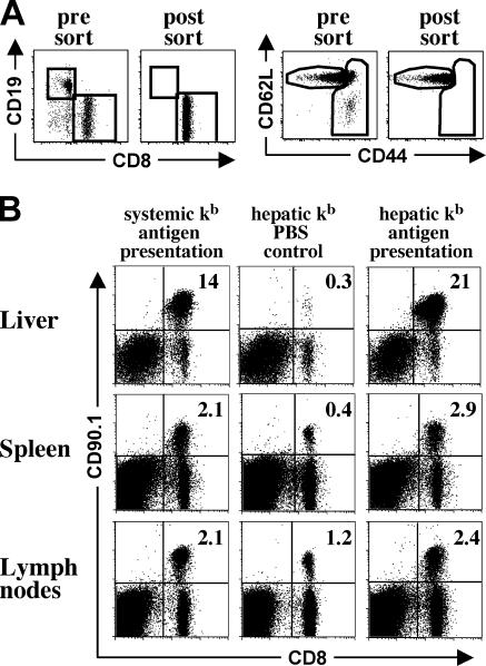
In vivo expansion of naive OT-I T cells after hepatic and systemic antigen presentation. Naive OT-I cells for adoptive transfer were obtained by magnetic bead depletion, followed by FACS sorting for transgenic T cells with a naive phenotype. (A) Purity and activation status of adoptively transferred OT-I T cells before and after flow cytometric cell sorting. (B) Expansion of adoptively transferred OT-I T cells detected in livers, spleens, and peripheral lymph nodes of transplant recipients. For restricted hepatic kb antigen presentation, livers from [bm8→B6] bone marrow chimeras were transplanted into bm8 recipients. Systemic kb antigen presentation was achieved by transplanting livers from [B6→B6] bone marrow chimeras into B6 recipients. 4 wk after transplantation, 5 × 10⁶ naive OT-I cells (CD90.1 background) were adoptively transferred and the animals received three i.p. injections of either SIINFEKL peptide or PBS (hepatic kb PBS control). 4 d after the initial antigen contact, transplant recipients were killed and the number of OT-I cells was assessed by flow cytometry. Data are representative of three independent experiments with three mice per group, and sorted and unsorted OT-I cells were used for adoptive transfer without significant differences in the extent of prolif-eration. This shows that antigen presentation in the transplanted liver can cause extensive clonal expansion.

