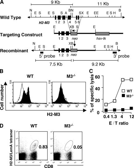
Figure 1.
Generation and characterization of M3−/− mice. (A) Genomic configuration of the H2-M3 locus before (top) and after (bottom) homologous recombination with the targeting construct (middle). Exons (black boxes), lengths of diagnostic restriction fragments, and the probes used for Southern analysis are shown. An arrow indicates the transcription orientation of the neo gene. B, BglII; N, NheI; E, EcoRI; S, SacI; A, ApaI; H, HindIII; X, XbaI. (B) Splenocytes from WT and M3−/− mice were incubated overnight with (filled histogram) or without (open histogram) 10 μM LemA peptide. Cells were stained with an anti–H2-M3 mAb 130, followed by FITC-anti–hamster IgG. (C) H2-M3–specific · CTL clone 4E3 was incubated with LPS blasts from WT and M3−/− during a 51Cr release assay. The effector–target ratio is indicated in the figure. (D) Identification of LM-specific H2-M3–restricted T cells after i.v. infection with LM. Peripheral blood lymphocytes were isolated from LM-infected M3−/− mice (N6) or WT controls on day 7 after infection. Cells were stained with FITC–anti-CD8α and PE-H2-M3/LemA-tetramer. CD8+ tetramer+ T cells are highlighted by the gate, and the proportion of gated cells is indicated.

